
Tendons and wounds share something frustrating: both heal slowly, and both frequently disappoint. Tendons are notoriously poor healers because of their limited blood supply and low cellular turnover. Wounds in older adults, diabetic patients, or anyone with compromised circulation can stall for weeks or months – becoming chronic wounds that standard care genuinely struggles to resolve.
Red light therapy addresses both through the same underlying mechanism, and the evidence, while different in strength between the two conditions, is solid enough that physical therapists, sports medicine physicians, and wound care specialists are increasingly incorporating it into clinical practice.
Here’s what the research actually supports, where the gaps are, and what a practical protocol looks like for each.
Why Tendons Are So Hard to Heal
Tendons connect muscles to bones. They’re built for tension – dense, organized cables of type I collagen that transmit enormous mechanical force. But that same density works against them in injury: tendons receive minimal direct blood supply, rely heavily on diffusion for oxygen and nutrients, and have very low metabolic activity under normal conditions. When damaged, cellular repair is slow.
Tendinopathy – a term that covers both acute tendonitis (inflammatory) and chronic tendinosis (degenerative) – is among the most common musculoskeletal complaints seen in clinical practice. Achilles tendinopathy, rotator cuff tendinitis, patellar tendinopathy (jumper’s knee), and lateral epicondylitis (tennis elbow) together affect millions of people annually. They share a common frustration: even with rest, physical therapy, and anti-inflammatory treatment, recovery can take months and reinjury is really common.
Standard-of-care approaches – eccentric loading exercises, NSAIDs, and in refractory cases, corticosteroid injections or surgery – manage symptoms but don’t directly address the biology of tendon repair. PBM does, at least in principle, and the research is bearing that out.
How Red Light Therapy Acts on Tendon Tissue
The mechanism begins, as always, with mitochondrial stimulation. But in tendon tissue, several downstream effects are particularly relevant.
Fibroblast activation and collagen synthesis – Tendons depend on fibroblasts (specifically tenocytes) for collagen production and matrix remodeling. PBM has been shown to stimulate fibroblast proliferation and upregulate type I collagen synthesis – the structural protein that gives tendons their tensile strength. A 2024 in vitro study published in Lasers in Medical Science (Cárdenas-Sandoval et al.) confirmed that LLLT increased type I collagen synthesis in early-stage fibroblast healing, with implications for both tendon and ligament repair.
Anti-inflammatory modulation – In acute tendonitis, PBM can downregulate pro-inflammatory cytokines (IL-1β, TNF-α) and reduce prostaglandin E2, reducing pain and swelling without suppressing the repair signals needed for healing. And honestly, that’s a meaningful advantage over NSAIDs in the recovery context.
Collagen remodeling – Chronic tendinopathy is characterized not just by inflammation but by disorganized collagen architecture – fibers laid down haphazardly rather than in the aligned bundles that give tendons their strength. PBM appears to support more organized collagen deposition, addressing the tissue quality problem that explains why many tendinopathies never fully resolve even after pain settles.
Nitric oxide and microcirculation – Near-infrared light triggers nitric oxide release, improving microvascular perfusion in poorly vascularized tendon tissue. Better local circulation is what makes more oxygen and growth factors available at the site of damage.
Nerve modulation – PBM can reduce substance P and other neuropeptides involved in peripheral sensitization, which helps explain why pain relief often precedes measurable structural changes. At the same time, it can be quite useful in chronic tendinopathy where neural sensitization has become part of the pain picture.
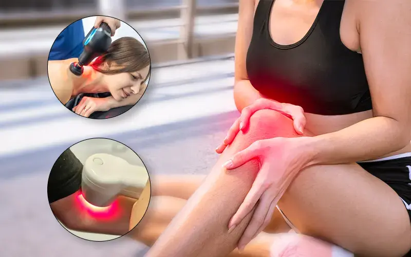
The Evidence for Tendonitis: Condition by Condition
Achilles Tendinopathy
This is the most studied tendon application for PBM – and it produces some of the clearest positive results. A widely cited 2008 randomized controlled trial by Stergioulas et al. in the American Journal of Sports Medicine found that low-level laser therapy combined with eccentric exercises produced significantly greater pain reduction and return to sport compared to eccentric exercises plus placebo laser in recreational athletes with chronic Achilles tendinopathy. And honestly, the key finding here is straightforward: PBM accelerated the benefit of the exercise protocol, getting patients to meaningful pain reduction faster.
A 2021 systematic review and meta-analysis in BMC Sports Science, Medicine and Rehabilitation (Tripodi et al.) examined PBM across tendinopathies and found real evidence of benefit for pain reduction – though it noted that heterogeneity in protocols (wavelength, dose, treatment frequency) made direct comparisons quite difficult, and called for more standardized trial designs. The biological signals are consistent; the protocol question is still being optimized.
A 2025 study (Lim et al., International Journal of Molecular Sciences) examined LED-mediated PBM in a murine tendon healing model, finding that photobiomodulation promoted collagen fiber organization and reduced inflammatory cell infiltration. That’s direct structural evidence supporting what the clinical pain data suggest.
Rotator Cuff Tendinopathy
The evidence here is meaningful but more mixed. A study in Lasers in Medical Science compared therapeutic ultrasound with LED photobiomodulation (850 nm infrared and 640 nm red) in 75 patients with shoulder tendinopathy aged 45–70. All PBM groups showed improvements in pain, range of motion, and quality of life, with the combined approaches showing advantages over either treatment alone. A 2025 study in Wound repair and regeneration examined PBM combined with exercise-based rehabilitation for rotator cuff pathology, finding improved pain and functional recovery outcomes compared to exercise alone.
The rotator cuff is a deep structure, and device penetration matters here. Near-infrared wavelengths (808–850 nm) are necessary to reach rotator cuff tissue meaningfully – surface-level red light alone isn’t sufficient for this application.
Lateral Epicondylitis (Tennis Elbow)
Multiple randomized trials have evaluated LLLT for lateral epicondylitis with generally positive results. A 2024 comparative RCT published in Rheumatology International found that high-intensity laser therapy was comparable to extracorporeal shock wave therapy in lateral epicondylitis for pain and functional outcomes. PBM is consistently cited in systematic reviews of conservative management options for tennis elbow, with a reasonable evidence base for short-to-medium term pain reduction.
Patellar Tendinopathy (Jumper’s Knee)
A 2022 BMJ Open systematic review analyzing 18 randomized trials found that low-level laser therapy (LLLT) consistently improved pain and function in people with lower extremity tendinopathy and plantar fasciitis. The greatest benefits were observed when guideline-recommended laser doses were used, supporting LLLT as a safe, non-invasive treatment option with no reported adverse events.
The Honest Picture for Tendonitis
PBM is best understood as an adjunct to, not a replacement for, active rehabilitation. Eccentric loading exercises remain the evidence-based cornerstone of tendinopathy management. What PBM appears to do is accelerate the pain reduction that allows patients to engage with exercise earlier and more effectively, and potentially improve the quality of tendon repair over time. It won’t fix a tendinopathy by itself if the mechanical loading problem isn’t addressed.
“Red light therapy is most effective for tendinopathy when it’s part of a broader rehabilitation plan,” says Dr. Michael Hamblin of Harvard Medical School, a leading PBM researcher. “The biology supports it as an accelerant of the healing process – not a substitute for the physical work of tendon remodeling.”
Red Light Therapy for Wound Healing: One of the Strongest Evidence Bases in PBM
Wound healing is where photobiomodulation has some of its most compelling and mature clinical evidence. The research spans decades, multiple wound types, and numerous rigorous systematic reviews – and the picture that emerges is consistently positive, particularly for wounds that are slow to heal under standard care.
How PBM Acts on the Four Stages of Wound Healing
Wound healing proceeds through four overlapping phases: hemostasis, inflammation, proliferation, and remodeling. PBM has documented effects across three of these:
Inflammation phase – PBM modulates the inflammatory response without suppressing it completely. It reduces excessive pro-inflammatory cytokines that can prolong the inflammatory phase and delay progression to tissue rebuilding, while preserving the antimicrobial functions that make initial inflammation necessary.
Proliferation phase – This is where PBM’s effects are most pronounced. It stimulates fibroblast proliferation and migration into the wound, upregulates collagen synthesis, promotes keratinocyte activity (re-epithelialization – the process of new skin growing across the wound), and stimulates angiogenesis (formation of new blood vessels). All four are essential for effective tissue rebuilding.
Remodeling phase – PBM influences the quality and organization of scar tissue. RCTs in post-surgical patients show PBM reduces scar erythema, improves scar elasticity, and improves objective scar quality scores through modulation of MMP expression and reduction of excess myofibroblast activity.
Chronic Wounds: Diabetic Ulcers and Pressure Wounds
This is where the clinical evidence is strongest and the clinical need is most acute. Diabetic foot ulcers are notoriously difficult to heal – impaired circulation, neuropathy, metabolic dysfunction, and chronic hyperinflammation combine to create a wound environment where standard care frequently produces inadequate results. Amputation risk is real.
Multiple RCTs in diabetic wound healing show 40–50% faster healing times with PBM adjunct therapy compared to standard care alone. The mechanism is particularly relevant in the diabetic context: PBM restores mitochondrial function in metabolically compromised diabetic cells, increases nitric oxide-mediated perfusion in poorly vascularized tissue, and reduces the chronic hyperinflammatory state that impairs healing.
A 2024 systematic review and meta-analysis in Cureus examining LLLT specifically for skin wounds confirmed significant improvements in wound healing rates and pain reduction, covering 670 wounds across multiple wound types including diabetic ulcers, surgical wounds, and burn wounds.
NASA research conducted in the 1990s – originally aimed at helping astronauts heal in zero gravity – established near-infrared light as an effective wound healing accelerant and helped build the foundation for the clinical evidence that followed.
Acute and Surgical Wounds
For acute wounds – surgical incisions, minor burns, donor site wounds – the evidence is positive but more modest than for chronic wounds. Several small RCTs show faster epithelialization, reduced pain, and improved comfort with PBM compared to sham.
The benefit in acute wounds is real but less dramatic than in chronic wounds, because acute wounds in otherwise healthy tissue heal reasonably well with standard care anyway. The gap that PBM most effectively closes is in compromised healing: elderly patients, diabetics, immunosuppressed individuals, or wounds in poorly vascularized tissue.
A 2025 consensus paper from the Journal of the American Academy of Dermatology on evidence-based clinical applications of PBM specifically included wound healing as one of the most robustly supported indications, citing evidence across diabetic ulcers, venous leg ulcers, surgical wounds, and oral mucositis.
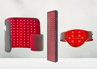
Wavelengths and Protocols That Actually Matter
For both tendonitis and wound healing, the right red light therapy wavelength depends on target tissue depth.
660 nm red light – The primary wavelength for wound healing at the skin surface. Directly stimulates keratinocytes, fibroblasts, and superficial tissue repair. Best for surface wounds, epithelialization, and scar remodeling.
808–850 nm near-infrared – Better penetration for deep tissue targets. Preferred for tendon structures (particularly deeper tendons like the rotator cuff), and for wounds with significant tissue depth or in poorly vascularized tissue where deeper penetration is needed to reach affected cells.
Devices combining both wavelengths are often most effective for wounds with both superficial and deep components, and for tendons where you want both anti-inflammatory surface effects and deeper tissue stimulation.
Practical protocol framework:
For tendonitis:
- 10–20 minutes per session directly over the tendon
- 3–5 sessions per week
- 4–8 weeks minimum before assessing results
- Near-infrared (808–850 nm) for deeper tendons; combined red/NIR for Achilles and surface structures
- Used alongside, not instead of, your rehabilitation program
For wound healing:
- 10–20 minutes per session over the wound area
- Daily treatment is supported in chronic wound research; every other day for acute wounds
- Begin treatment after the wound is closed or in the granulation phase – not on actively bleeding wounds
- 660 nm for surface wounds; 850 nm or combined for deeper or chronic wounds
- For diabetic foot ulcers specifically: daily treatment for 4–8 weeks is most consistently studied
On dose: Total energy delivered per session (J/cm²) matters as much as time. The biphasic dose-response curve applies here – too little has no effect, the right amount helps, and too much can paradoxically inhibit healing. Most wound healing studies use 2–6 J/cm² per session; tendinopathy studies vary more widely (4–20 J/cm²). Following manufacturer dosing guidelines and erring toward the lower end initially is appropriate for home use.
What to Realistically Expect
Tendonitis: Most clinical trials show meaningful pain reduction within 4–6 weeks of consistent PBM combined with appropriate exercise rehabilitation. You’re unlikely to notice a dramatic change after one or two sessions. What tends to happen is a gradual reduction in morning stiffness and post-activity pain, with improved ability to engage in the eccentric loading exercises that do the structural repair work.
Wound healing: Response depends heavily on wound type and the patient’s baseline health. In diabetic foot ulcers and chronic wounds, the evidence supports faster healing times and improved closure rates – but this is measured in weeks of treatment, not days. For acute surgical wounds in healthy patients, improvement in comfort and closure speed is measurable but more modest.
Neither application is a miracle – PBM works with your body’s healing biology, not instead of it. Chronic wounds still need proper wound care. Tendinopathy still needs mechanical rehabilitation. What PBM adds is meaningful acceleration and improvement of tissue quality – and for slow-healing conditions, that’s a genuinely valuable contribution.
When Not to Use Red Light Therapy on a Wound
Most wounds and tendon conditions are appropriate for home PBM use with reasonable care. A few situations warrant caution or medical involvement first:
- Active infection in or around the wound – PBM should not be used over infected tissue without medical supervision. Treat the infection first.
- Open wounds with active bleeding – wait until hemostasis is established and the wound has entered the inflammatory/granulation phase
- Wounds near implanted electronics – avoid direct exposure over pacemakers, neurostimulators, or similar implanted devices
- Cancer patients with active skin involvement – do not treat directly over known or suspected malignant tissue
- Very deep or tunneling wounds – these require professional wound care management regardless of adjunct therapies
Frequently Asked Questions
Does red light therapy help tendonitis?
A: Yes – clinical evidence supports PBM for pain reduction and functional improvement in multiple tendinopathies including Achilles, rotator cuff, lateral epicondylitis, and patellar tendinopathy. It works best as an adjunct to active rehabilitation, not a standalone treatment. Expect meaningful results over 4–8 weeks of consistent use.
How long does it take for red light therapy to work on tendons?
A: Most studies show measurable pain reduction within 4–6 weeks of consistent treatment (3–5 sessions per week). Some people notice reduced stiffness sooner. Structural improvement in tissue quality takes longer – chronic tendinopathies that have been present for months won’t resolve in two weeks regardless of treatment.
Can red light therapy heal wounds faster?
A: It can meaningfully accelerate healing in chronic wounds and compromised healing conditions. Meta-analyses show 38–50% faster closure in diabetic foot ulcers with PBM adjunct therapy. For acute wounds in healthy patients, the benefit is real but more modest. PBM doesn’t replace proper wound care – it enhances it.
What wavelength is best for wound healing?
A: 660 nm red light for surface wounds and epithelialization. 808–850 nm near-infrared for deeper wounds or in poorly vascularized tissue. Devices combining both wavelengths cover the full depth spectrum.
Can I use red light therapy on an open wound?
A: Wait until active bleeding has stopped and the wound is in the inflammatory or early granulation phase. Don’t apply directly over infected tissue. For chronic wounds like diabetic ulcers, PBM is well studied when applied to wounds that are open but not actively bleeding or acutely infected.
Is red light therapy used by physical therapists and doctors for tendonitis?
A: Yes. PBM is increasingly used in physical therapy, sports medicine, and orthopedic settings as an adjunct for tendinopathy management. Cold laser devices are standard equipment in many PT clinics for this application.
The Bottom Line
Of all the musculoskeletal and tissue repair applications of red light therapy, wound healing and tendinopathy sit among the best-supported. Wound healing – particularly for chronic wounds like diabetic ulcers – has decades of clinical evidence, multiple systematic reviews, and a clear mechanistic basis. Tendinopathy evidence is meaningful and growing, with the clearest support for Achilles, patellar, and rotator cuff presentations.
Neither is a replacement for proper clinical management. But for conditions that routinely frustrate both patients and clinicians under standard care, having a well-evidenced, non-invasive, low-risk adjunct that demonstrably accelerates repair is worth taking seriously. The biology is sound, the protocols are improving, and the clinical uptake is following the evidence.